
Leptographium wageneri
Leptographium wageneri
Leptographium wageneri is a specialized ascomycete fungus belonging to the order Ophiostomatales. It is the causal agent of black stain root disease, a serious infectious vascular wilt affecting various conifer species.
What the section contains
Leptographium wageneri
This microorganism is classified as a mycopathogen that attacks the vascular system of trees. It is taxonomically related to fungi that are often transmitted by specific insect vectors, particularly bark beetles and weevils.
The fungus mycelium within the plant tissues is characterized by dark, almost black pigmentation, which causes the internal wood to become stained. It produces conidia in specialized structures known as conidiophores with dark heads.
The species is biologically adapted to live within the xylem of conifers, where it proliferates and progressively blocks the transport of water and nutrients to the crown of the tree.
Microscopic examination of affected wood samples allows for precise identification of the pathogen based on the morphological characteristics of its spores and hyphae, distinguishing it from other species of the genus Leptographium.
The primary hosts for Leptographium wageneri are species within the Pinus genus, as well as certain spruce and Douglas-fir species. Mature forest stands are particularly susceptible to this infection.
The fungus attacks the root system and the lower stem, causing severe physiological disruption. Damage to the vascular tissues hinders normal upward water flow, which inevitably leads to the gradual decline of the tree.
The economic impact manifests as a loss of wood quality due to staining and necrosis. Ultimately, severe infection leads to tree mortality, causing significant damage to forestry and timber production.
The pathogen can spread not only via insects but also through root contacts between neighboring trees. This process facilitates the formation of infection centers where the disease moves from one tree to another.
Young trees often succumb to the disease more rapidly than mature ones, which seriously hampers the natural regeneration of forest stands in areas where the fungus is endemic.
Early symptoms of infection include a change in needle color from healthy green to a dull, faded green, followed by gradual yellowing and browning. The crown becomes thin, and needle drop is observed.
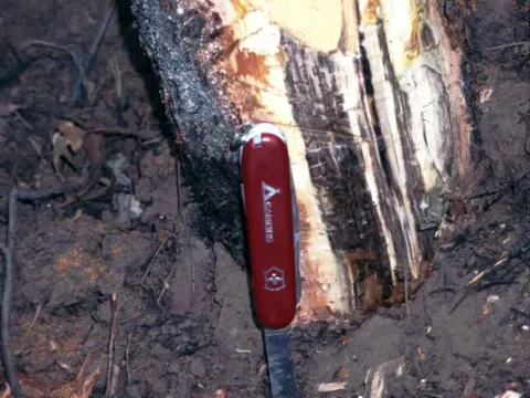
Cross-sections of affected wood, particularly near the base of the stem, reveal distinct radial dark streaks or sectors. These necrotic changes result from the tracheids being filled with fungal mycelium.
Reduced height and diameter growth are important diagnostic indicators that frequently precede the total death of the tree. The tree gradually loses its vitality over several growing seasons.
While increased resin flow may be observed in some cases, severe vascular wilt often results in a total cessation of resin production due to the death of living cambium and phloem cells.
A definitive diagnosis is established through laboratory analysis, which includes isolating the fungus in pure culture on nutrient media and confirming its pathogenic characteristics.
The primary method for management is conducting sanitation cuttings within forest stands. Removing infected trees, including their root systems, helps reduce the fungal population and prevents further spread.
It is crucial to restrict the movement of infested timber from infected areas to healthy forest plots. Controlling the transport of bark-covered logs is a critical prophylactic measure.
Application of insecticides to manage insect vectors, such as bark beetles and weevils, helps lower the risk of fungal spore transmission to healthy trees during the insects' flight periods.
The selection and planting of conifer species resistant to this fungus represent a promising strategy for reforestation. Genetically adapted trees are significantly better at resisting the infection.
In forest nurseries, maintaining high standards of silvicultural practice, providing optimal growth conditions, and avoiding root damage during transplantation are essential to prevent creating entry points for the pathogen.