
Hymenochaete agglutinans
Hymenochaete agglutinans
Description
How to identify
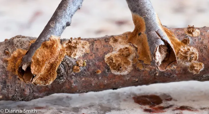
Hymenochaete agglutinans is a parasitic fungus belonging to the kingdom Fungi, phylum Basidiomycota. It is known primarily for its unique ability to colonize and physically bind together twigs and branches of various woody plants.
The fungus produces thin, leathery fruit bodies that appear as brownish crusts on the bark of its host. These fruit bodies are often velutinous (velvety) in texture and are characteristic of the Hymenochaetaceae family.
Microscopic examination reveals the presence of setae, which are diagnostic spine-like structures found within the hymenium. These structures assist in identifying the species under laboratory conditions.
The organism thrives in high-humidity environments, spreading via basidiospores that are released into the air and travel to new susceptible hosts, particularly through bark wounds or fissures.
Biology of the fungus involves the development of a dense mycelial mat that essentially "glues" the vegetation together, which is the specific behavior that gives the organism its scientific name.
What it damages
This pathogen targets a wide variety of deciduous trees and shrubs, including fruit trees like apples and pears, as well as several ornamental species found in parks and nurseries.
The damage is caused by the fungus disrupting the vascular system and cambium layer of the branches. By gluing branches together, it inhibits natural movement and causes localized tissue death.
As the infection progresses, the foliage on the affected branches begins to wither and die, leading to significant dieback of the crown and, in severe cases, the death of the entire tree.
Young trees in nurseries are particularly vulnerable to Hymenochaete agglutinans, where dense spacing facilitates the rapid spread of the fungus from one plant to another.
The aesthetic and economic value of the plants is severely diminished, requiring intensive intervention, including the pruning or removal of the infected plant material.
Signs of infestation
The most distinctive sign is the binding of adjacent twigs, creating a visible structure where branches appear to be stuck together by a crusty, leathery material.
Affected bark shows signs of necrosis; it may become discolored, cracked, or sunken as the fungus penetrates deeper into the woody tissue, effectively cutting off nutrient transport.
In the late stages of infection, the host displays premature leaf senescence, yellowing, and falling, as the branch distal to the fungal colony loses its supply of water and essential nutrients.
During the growing season, especially after rainfall, the fruit bodies of the fungus become more pronounced, appearing as rusty-brown, flat growths on the bark surface.
Removing a section of the affected bark reveals a whitish to yellow fungal mycelium underneath, which indicates an established infection that has begun to decay the host's inner tissues.
Control measures
The primary control measure is aggressive sanitation, which includes pruning and destroying all infected branches. It is crucial to cut well into healthy tissue to ensure the complete removal of the fungus.
Cultural practices such as pruning for air circulation are essential. By reducing the density of the crown, the environment becomes less favorable for the moisture-dependent fungal growth.
All pruning tools must be disinfected between cuts to prevent cross-contamination. Any accidental wounds or pruning cuts should be sealed to prevent spores from entering the tree.
Prophylactic treatments using copper-based fungicides can be applied in early spring before bud break to provide a protective barrier against potential spore germination.
Maintaining overall tree vigor through proper fertilization and watering helps the host plant build natural defenses, which can help limit the spread of the pathogen if infection occurs.
Causes diseases · 1
Discussion
No discussions yet — be the first.