
Heligmosomoides
Heligmosomoides
Description
How to identify
Heligmosomoides is a genus of parasitic nematodes belonging to the order Strongylida and the family Heligmosomidae, commonly studied in parasitological research.
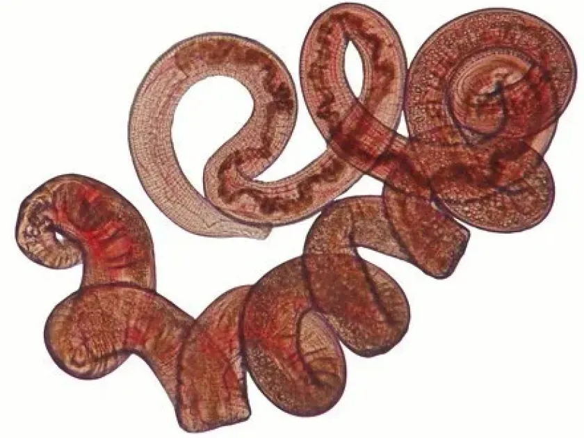
These worms are characterized by a spirally coiled body and a cuticle featuring longitudinal ridges, which help them attach to the host's intestinal lining.
The most well-known species, Heligmosomoides polygyrus, serves as a crucial model organism for studying intestinal helminth infections and host immune responses.
Morphological identification requires microscopic examination, specifically analyzing the reproductive structures and the bursal morphology of male specimens.
Diagnosis in live animals is typically achieved through fecal egg counts (FEC), identifying the characteristic oval-shaped eggs within the host's excrement.
What it damages
Heligmosomoides primarily affects laboratory and wild rodents, living in the lumen of the small intestine and invading the intestinal mucosa.
The presence of these parasites causes chronic inflammation, resulting in malabsorption of nutrients and significant weight loss in the affected animals.
Severe infestations can lead to stunted growth, reduced vitality, and an overall weakened immune system, making hosts susceptible to secondary pathogens.
In research facilities, these infestations can skew experimental data and increase the costs associated with animal care and long-term health management.
The cumulative damage to the intestinal epithelium may cause lasting physiological distress, impacting the overall welfare of the rodent population.
When it appears
The life cycle of Heligmosomoides is direct, requiring no intermediate host, which allows for rapid spread within confined rodent colonies.
Eggs shed in the feces develop into infective larvae in the environment, provided there is sufficient moisture and appropriate temperatures.
Warm and humid environments significantly accelerate the development of larvae, increasing the risk of infection during specific seasons.
In indoor facilities, improper sanitation or damp bedding can allow larvae to persist as an ongoing source of re-infection for the population.
High population density within cages exacerbates the speed of transmission, leading to rapid outbreak cycles if not monitored strictly.
Signs of infestation
Clinical signs of an infestation include noticeable weight loss, despite the animal maintaining a normal or even increased appetite.
Affected rodents often display a dull or ruffled coat, reduced activity levels, and general lethargy compared to healthy individuals.
Gastrointestinal distress is common, frequently manifesting as diarrhea, which can lead to dehydration and further metabolic complications.
Post-mortem examination often reveals localized mucosal thickening or nodules in the duodenum and jejunum, where the parasites attach.
Confirming the presence of the parasite requires fecal analysis to detect eggs, which is the standard diagnostic indicator for Heligmosomoides.
Control measures
The primary control strategy involves systematic deworming programs using anthelmintics effective against intestinal nematodes.
Strict sanitation protocols, such as regular cage cleaning and the removal of soiled bedding, are essential to break the transmission cycle.
Quarantine and screening of newly introduced animals are critical steps to prevent the accidental entry of parasites into a healthy colony.
Monitoring the health status of animals via routine diagnostic testing allows for early intervention before an infestation becomes widespread.
Combining pharmaceutical treatments with rigorous environmental management provides the most sustainable defense against this persistent parasite.
Taxonomy
- Latin name
- Heligmosomoides
- Order
- Nematodes
- Family
- Heligmosomidae
- EPPO code
- HLGSSP
Discussion
No discussions yet — be the first.